 Türkçe
 русский
 Deutsch
Oral Diagnosis and Radiology
Oral Diagnosis and Radiology
The mission of oral diagnosis and radiology department is to examine oral cavity through the x-ray. After detailed analysis, dentist looks into the patient’s x-ray results and diagnose.
Also preliminary diagnosis is done for the purpose of understanding of oral cavity diseases which will occur in future.
Diagnosis and Therapy
The subject of the department of oral diagnosis and radiology is to apply correct method and therapy for oral health by using screening systems. Detailed oral exam is done in our polyclinic after medical records of patients are taken. A proper plan is prepared for therapy with the assist of screening systems after examination.
Oral Diagnosis and Radiology Methods
Radiology
Oral cavity screening systems facilitate diagnosis of jaw bone diseases and assist dentists during the examination process. With the ultimate technology, most common used oral cavity screening systems are; intra oral camera, panoramic x-ray, periapical x-ray, digital x-ray, computed tomography.
Intra oral camera
Entire tooth can be visualized to see the clear picture of tooth roots, fillings, bridges, crowns by patient and dentist. Through the system diagnosis can facilitate and detailed examination can be done for tooth in the oral cavity.
Panoramic x-ray
X-rays are the pictures of the teeth, bones and soft tissues around them to help find out the problems of the teeth, mouth, and jaw. X-ray pictures can show cavities, hidden dental structures such as wisdom teeth, upper and lower jaw with tissues.
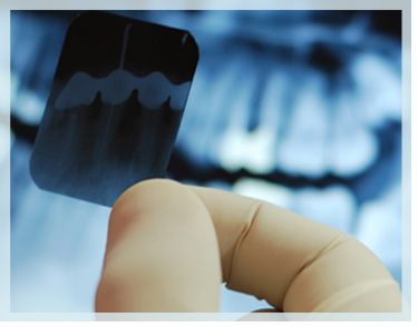
Periapical x-ray
This type of x-ray takes a full, very detailed tooth picture from the very top of the tooth (crown) to the very tip of the root. Periapical x-rays are usually taken when you are having symptoms with a specific tooth and intra oral disease. This type of x-ray is also used for adjacent tooth and its tissue to take their picture.
Computed tomography (CT)
X-ray Computed Tomography is a technique for visualizing interior features which can not be visualized by other techniques, to obtain digital information on their 3-D geometries and properties. This method allows to get cross-sectional picture of the part of body which is affected by disease and is used for patients who will be applied multiple implants.
Private Hurma Dental Health Polyclinic
Implant Placement Center - TURKEY